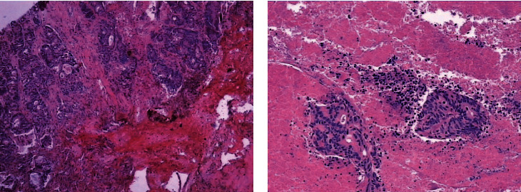
Figure 1.

H&E staining for the tissues at different time points. (a) H&E staining of tissues before radiofrequency ablation (magnification: 10 × 5). (b) H&E staining of tissues from biopsy immediately after radiofrequency ablation (magnification: 10 × 5).
