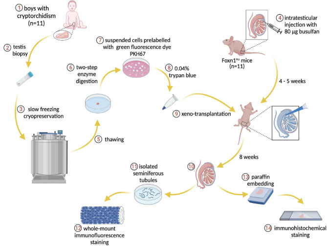
Figure 1.
Schematic overview of the experiment. Donor testis biopsies from infant boys with cryptorchidism underwent a frozen-thawed procedure and were dissociated through two-step enzymatic digestion. The suspended cells were prelabeled with PKH67 for tracing human cells within the recipient mouse seminiferous tubules post-transplantation. The endogenous spermatogenesis of recipient mice was eliminated by busulfan treatment. The human testicular cells were injected into the mouse tubules and the transplantation process was visible via trypan blue. Eight weeks later, the testes were harvested for immunostaining and further analysis.

