FIGURE 4.
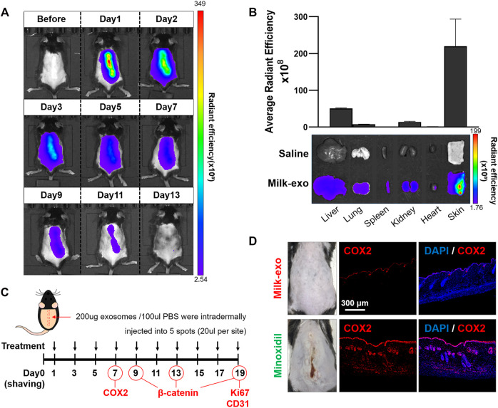
In vivo biodistribution of Milk-exo and expression of COX2 in skin tissue. (A) Real-time in vivo imaging of Cy5.5-NHS labeled exosome in C57BL/6 mice for 13 days after intradermal injection. The mice were analyzed at the indicated times after intradermal injection of 200 µg per 100 µL of exosomes. (B) Ex vivo imaging and Milk-exo quantification of the skin and major organs on day 2 after intradermal injection of labeled exosomes (n = 3). (C) Schedule of Milk-exo treatment (black arrows) and the date of histological analysis (COX2, β-catenin, Ki67 and CD31). (D) Representative immunostaining images of the expression of COX2 in mice skin tissues treated with Milk-exo and Minoxidil. C57BL/6 mice were sacrificed on day 7 after intradermal injection every 2 days. The nuclei (blue) were stained with DAPI, and Alexa Fluor® 647 conjugated secondary antibodies (red) were used for visualization of COX2.

