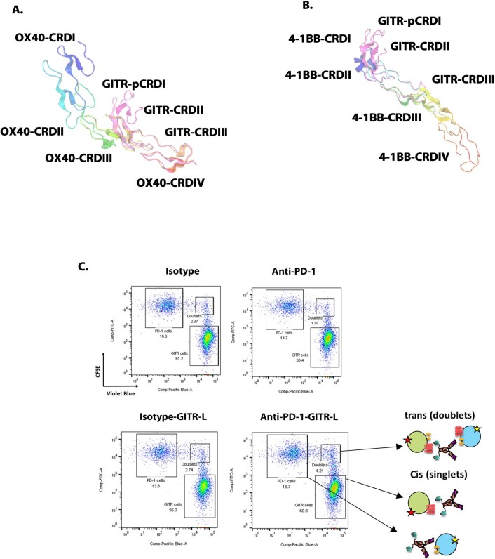
Extended Data Fig. 1. GITR structural alignment comparison with other TNFR members and flow cytometry cell bridging assay with the anti-PD-1-GITR-L bispecific.
(A and B) GITR (magenta) overlaid with OX40 (PDB: 2HEV, rainbow, RMS 1.3) (A) and 4-1BB (PDB: 6BWV, rainbow, RMS 1.1) (B). Note that CRDI of GITR (pCRD1GITR) is only partially resolved due to compositional heterogeneity. (C) A 1:1 combination of CFSE-labeled PD-1-HEK293 and Violet-blue-labeled GITR-HEK293 cells were treated with 2.5 µgs/ml of isotype control, anti-PD-1 mAb, isotype-GITR-L construct and anti-PD-1-GITR-L bispecific for 30 mins.

