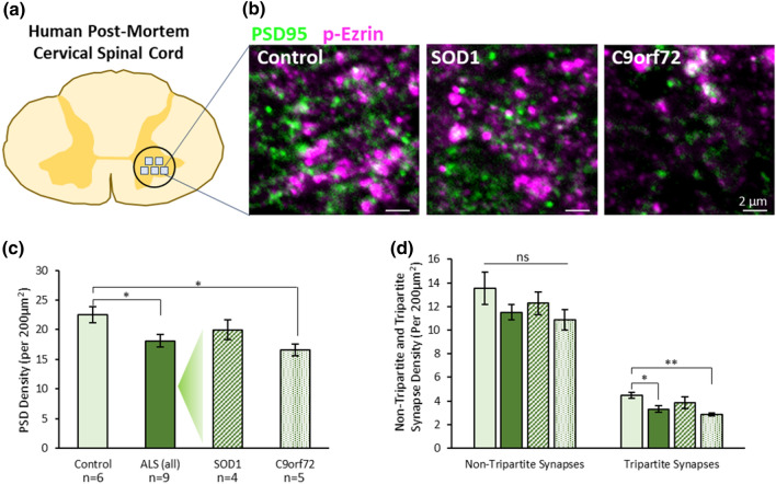
Fig. 4.
Tripartite Synapses are selectively lost in the cervical spinal cord of human ALS cases. a Diagram of human cervical spinal cord and location of image sampling. b Example images of synapses (PSD95—green) and astrocytic processes (p-Ezrin—magenta) in human spinal cord tissue. c Graph showing the total PSD density (PSD95 puncta) in controls, all ALS cases, and then SOD1 and C9orf72 cases separated. d Graph showing synapse density in patient groups when synapses are separated based on whether they are non-tripartite synapses or tripartite synapses, as determined by the association of PSD95 with p-Ezrin

