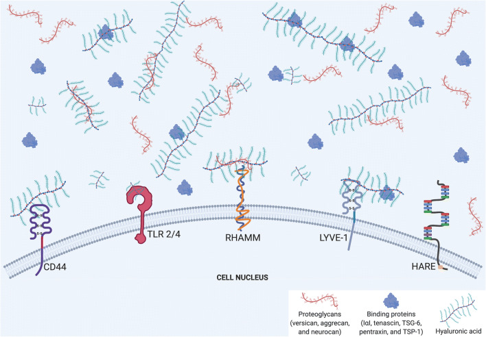
Fig. 1.
Schematic illustration of the interactions of extracellular hyaluronan (HA) and its receptors and binding proteins. This figure illustrates the common binding mechanisms for extracellular HA. The major HA receptors include CD44 and its homolog, LYVE-1, in addition to TLR 2/4, RHAMM, and HARE. These receptors are present at the cell surface, with extracellular HA binding with or without binding proteins (specified in the legend). The fact that HA and its binding proteins are located outside the cell indicates that HA is a major component of pericellular coats. The interaction between HA and its receptors can be preferential, as LMW-HA tends to bind TLRs 2/4, although some, like CD44, bind both LMW- and HMW-HA. The depiction of different sizes of HA corresponds to LMW and HMW variants, which are responsible for the dichotomous pro-inflammatory/pro-fibrotic effects to anti-inflammatory/anti-fibrotic effects, respectively. In addition, crosslinks between HA and various binding proteins, including proteoglycans and other hyaladherins, modifies HA’s effects. This schematic is a broad illustration and is not drawn to scale. Moreover, that five receptors are shown on one cell surface is for illustrative purposes only. Created with BioRender.com

