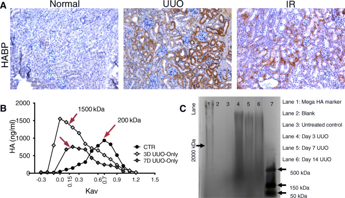
Fig. 2.
HA expression and molecular weight changes in normal and diseased mouse kidneys. A. Images of HA binding protein (HABP) staining of the cortex from control, 7-day UUO kidney and 7-day IR kidney show increased HA accumulation after UUO injury. Scale bars 50 μm. B. Plots of total incorporated [3H] in labeled HA samples were used to determine the relative MW of HA synthesized by control and 3D UUO kidneys (n ≥ 3 per condition) using Sephacryl S-1000 chromatography. Data were plotted as HA concentration versus the partition coefficient (Kav), showing an increase in HA size distribution in samples with UUO injury. C. The extracted HA from control and 3-, 7-, and 14-day untreated and treated UUO kidneys is shown on a 0.5% agarose gel electrophoresis, supporting the chromatography results

