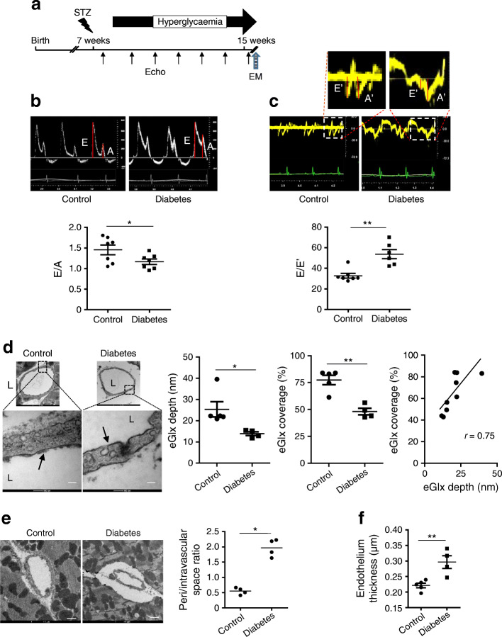
Fig. 1.
EGlx damage is associated with the development of DCM in FVB mice. (a) Diabetes was induced in FVB male mice by injection of low doses of STZ. The development of DCM was monitored with echocardiography for 9 weeks after STZ injection. (b, c) Representative pulsed wave Doppler images and reduced E/A ratio (b) and representative tissue Doppler images and increased E/E′ (c) in mice 9 weeks after STZ injection compared with control mice (n = 7 for control and 6 for diabetes; *p< 0.05 and **p< 0.01 [unpaired t test]). (d) Reduced eGlx depth and coverage in mice with DCM (n = 5; *p< 0.05 [unpaired t test]). Low- and high-magnification electron micrographs of capillaries from left ventricles of STZ-injected and control mice 9 weeks after STZ injection. Arrows show eGlx on top of endothelial cells. Scale bar, 100 nm. eGlx coverage (percentage of grid points at which eGlx depth is not less than 10 nm) is strongly positively associated with eGlx depth (n = 9; *p< 0.05 [Pearson r = 0.75]). (e, f) Increased peri/intravascular space ratio (area of perivascular space/area of intravascular space; n = 4; *p< 0.05 [unpaired t test]) (e) and endothelial thickness (n = 4 or 5; **p< 0.01 [unpaired t test]) (f) in the capillaries from diabetic vs non-diabetic hearts. Scale bar, 1 μm. Data are presented as mean ± SEM. EM, electron microscopy; L, capillary lumen

