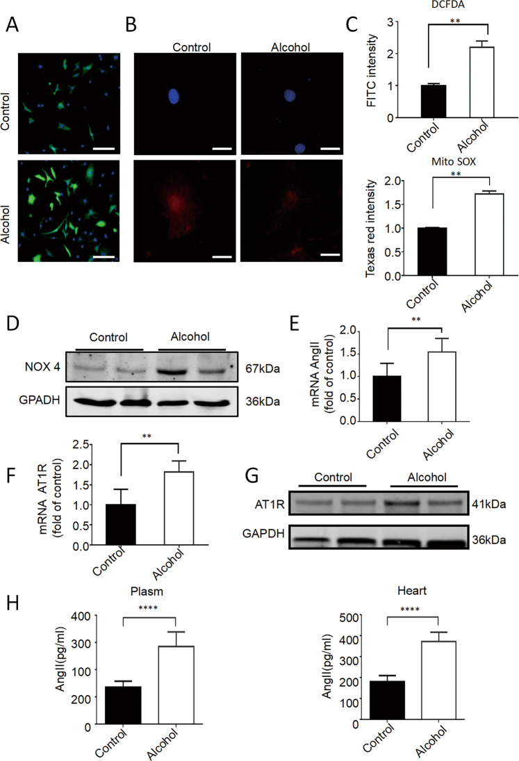
Fig. 4. Alcohol-induced ROS and AngII/AT1R activation in cardiomyocytes.
A–C Representative immunofluorescence images of CM-H2DCFDA (scale bar = 15 μm) and Texas red labeling (scale bar = 25 μm), and quantitative statistics showed that the level of the oxidative stress and mitochondrial oxidative stress significantly increased after hiPSC-CMs were treated with 100 mM alcohol for 2 h (n = 10). D Western blot showed that AT1R protein expression increased significantly after treatment with 100 mM ethanol for 24 h (n = 5). E RT-PCR showed that the expression of AngII and AT1R expression increased significantly after treatment with 100 mM alcohol for 24 h (n = 5). F RT-PCR showed that the expression of AngII and AT1R expression increased significantly after treatment with 100 mM alcohol for 24 h (n = 5). G Western blot showed that AT1R protein expression increased significantly after treatment with 100 mM alcohol for 24 h (n = 5). H Enzyme-linked immunosorbent assay showed that the level of AngII in the plasma and heart of mice increased after alcohol treatment (n = 6). Results are presented as means ± SEM (**P < 0.01; two-sided Student’s t-test or one-way analysis of variance).

