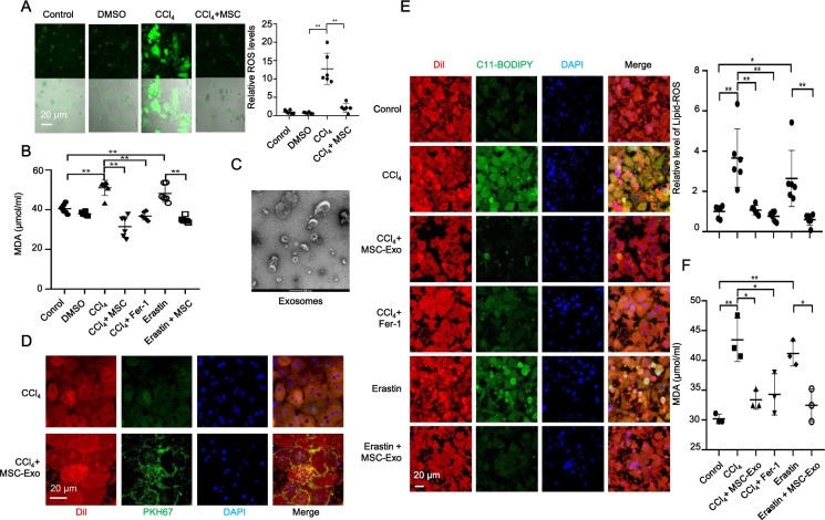
Fig. 4. MSC-Exo mediated the effect of MSCs against CCl4-induced ferroptosis in vitro.
A Relative ROS levels were measured by H2DCFDA staining in primary hepatocytes treated with control (PBS), dimethyl sulfoxide (DMSO), or 10 mM CCl4 for 48 h with or without MSCs coculture. B The increased MDA level induced by CCl4 was downregulated by MSCs coculture and Fer-1 treatment in primary hepatocytes. The increased MDA level induced by erastin was also downregulated by MSCs coculture in primary hepatocytes. C Refrigerated transmission electron microscopy was used to assess exosomes isolated from MSCs. D Exosomes collected from MSC-conditioned medium were labeled with PKH67 and incubated with CCl4-induced acute injured hepatocytes for 24 h. The cell membrane was stained with Dil. The nuclei were stained with DAPI. PKH67 lipid dye was detected in damaged hepatocytes treated with PKH67-labeled exosomes in the CCl4 group. E Isolated hepatocytes were incubated with C11-BODIPY581/791 and then analyzed by using laser confocal focus. Cell membrane was stained with Dil. The nuclei were stained with DAPI. C11-BODIPY581/591 staining showed a significant increase in lipid peroxidation in hepatocytes following CCl4 and erastin treatment, while MSC-Exo and Fer-1 downregulated the increased lipid peroxidation induced by CCl4 and erastin. F The increased MDA level induced by CCl4 and erastin were downregulated by MSC-Exo and Fer-1 treatments in primary hepatocytes. Significance was calculated by one-way ANOVA with Tukey’s post hoc test. *p < 0.05 or **p < 0.001 indicated a significant difference between groups.

