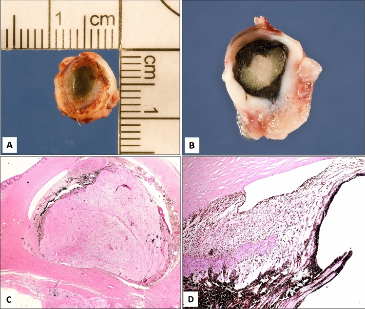
Figure 2.
(A,B) The gross appearance of a globe with severe microphthalmos. (C) Histopathological appearance of the globe with absent lens and dysgenesis of the anterior chamber angle structures and gliosis of the retina (original magnification × 12.5, hematoxylin and eosin). (D) Higher power of the anterior segment showing absent angle structures with uveal tissue adherent to the back of the cornea and obliterated narrow anterior chamber (original magnification × 100, hematoxylin and eosin).

