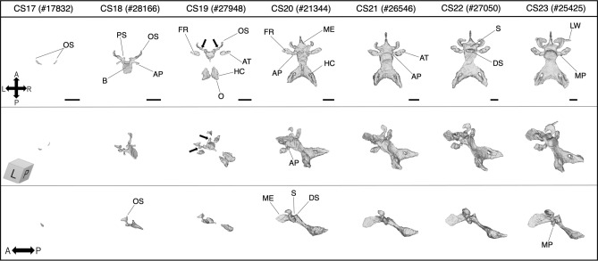
Figure 3.
The 3D models reconstructed using PCX-CT. Typical appearance of the sphenoid (including the occipital and the nasal septum) in each stage (black bar: 1 mm). Note that these are not necessarily corresponding to the findings recorded in Table 2 and Fig. 2 which are the results of HS observation. Arrows (CS19) indicate a structure that articulates with the orbitosphenoid and presphenoid, which is called “postorbital root of the orbital wing” or “hypochiasmatic alae” in previous reports. HS, histological section; PCX-CT, phase-contrast CT; AT, ala temporalis; AP, alar process; B, basisphenoid; DS, dorsum sella; FR, foramen rotundum; HC, hypoglossal canal; LW, lateral part of the lesser wing; ME, mesethmoid; MP, medial pterygoid process; O, occipital; OS, orbitosphenoid; PS, presphenoid; S, sella tunica.

