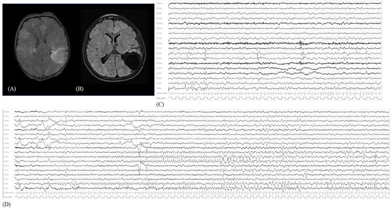
FIGURE 3.

(A) FLAIR‐weighted MRI with bilateral tubers, among which the most prominent over the left parietal lobe. (B) Postsurgical FLAIR sequence showing the parietal resection. (C) Wakefulness interictal video‐EEG with left parietal and vertex epileptiform abnormalities. (D) Video‐EEG recording of left parietal focal seizure, with rhythmic theta activity over C3–P3 and anterior vertex, evolving in focal spike and sharp waves discharge. EEG, electroencephalography; FLAIR, fluid attenuated inversion recovery; MRI, magnetic resonance imaging.
