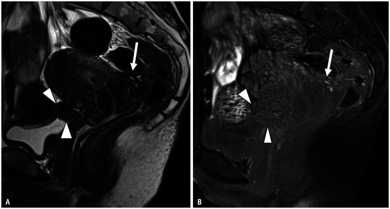
Fig. 11. Pelvic and bladder endometriosis in a 39-year-old female after hospital admission because of infertility.
A, B. Sagittal T2WI (A); Sagittal T1WI (B). Irregularly shaped nodules are observed at the level of the torus uterinus (arrows). Low signal intensity is observed on T2WI (A) and include hyperintense cysts on fat-suppressed T1WI (B). These findings indicate fibrosis caused by deep infiltrating endometriosis. The anterior surface of the rectum is strongly stretched to the torus uterinus. The anterior wall of the uterine cervix and lower uterine body show low signal intensity, probably reflecting fibrosis caused by adenomyosis. Continuing from uterine adenomyosis, soft tissue (arrowheads) with low signal intensity on T2WI continues to the bladder wall. Hyperintense foci on T1WI are visible at the mass periphery. Although focal bladder thickening resembles a bladder tumor, a hyperintense cyst on T1WI and the presence of adenomyosis and fibrous tissue around the lesion led to a diagnosis of endometriosis. WI = weighted image

