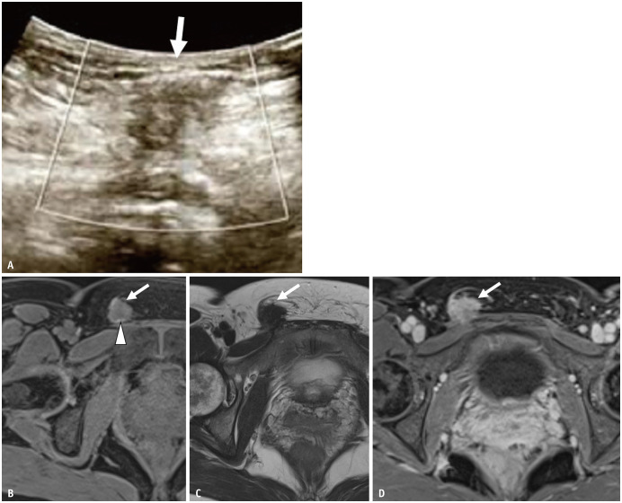
Fig. 12. A 46-year-old female with inguinal lesion.
The patient had undergone laparoscopic cystectomy for a left endometriotic cyst 10 years previously.
A. On ultrasound, the lesion (arrow) is visualized as a hypoechoic round nodule with an ill-defined border. B-D. Axial FS T1WI (B); axial T2WI (C); axial contrast-enhanced FS T1WI (D). The lesion (arrows) shows low signal intensity on both T1 and T2WI with an ill-defined border. There is a small high-intensity spot on FS T1WI (arrowhead). The mass is almost homogeneously enhanced. The patient received hormonal therapy, which reduced the lesion. FS = fat saturated, WI = weighted image

