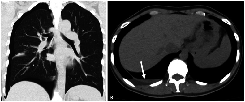
Fig. 13. A 45-year-old female with regularly recurring pneumothorax.
A, B. CT without contrast. Pneumothorax is found on the right side. A small amount of pleural effusion (arrow) is also observed. The fluid density is somewhat high. Hemothorax is suspected. During the operation, a small defect is observed in the diaphragm. It was thought to be the migration path of the endometrial tissue from the peritoneal cavity to the pleural cavity. On the basis of a pathological examination, endometriosis was diagnosed from the tissue around a small defect on the diaphragm.

