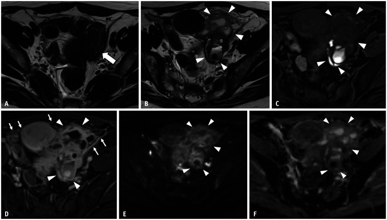
Fig. 2. Rupture of an ovarian endometriotic cyst in a 39-year-old female with abdominal pain and fever.
A. Axial T2WI 2 months before the rupture of the ovarian endometriotic cyst. A lesion with low signal intensity (arrow) is observed just beside the uterus. This is an ovarian endometriotic cyst with shading. B, C. MRI images obtained 2 months after (A) (B, axial T2WI; C, axial T1WI with fat saturation). On axial T2WI, the left ovarian endometriotic cyst (arrowheads) is now visualized as multiple loculated cysts with fluid–fluid levels and mixed signal intensities on T1 and T2WI, signifying a recent hemorrhage. The anterior wall of the cyst is irregular, and hyperintense fluid is observed around the cyst (arrowheads). These findings suggest rupture of an ovarian endometriotic cyst. D. Axial contrast-enhanced T1WI with fat saturation, irregularly thickened and enhanced cysts (arrowheads), and strong peritoneal enhancement (arrows) are observed. Tubo-ovarian abscess caused by infection after endometriotic cyst rupture was suggested and confirmed postoperatively. E, F. Axial DWI (b = 1000 s/mm2) (E); Axial apparent diffusion coefficient map (F). No area (arrowheads) shows restricted diffusion within the tumor, probably because the MRI image was obtained shortly after the rupture of the tumor. WI = weighted image

