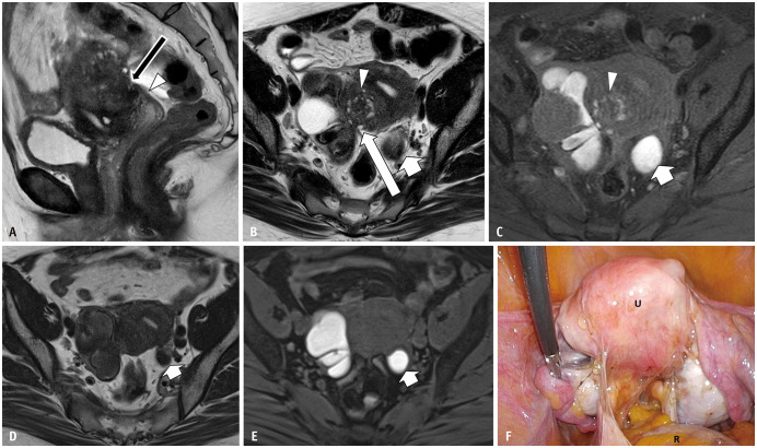
Fig. 3. Bilateral endometriotic cysts with Douglas obliteration in a 46-year-old female with dysmenorrhea and lower abdominal pain.
A-C. Sagittal T2WI (A); axial T2WI (B); axial T1WI (C) with fat saturation T2WI imaging shows an elevated posterior vaginal fornix (A, arrowhead), fibrotic plaque on the serosal surface of the uterus (A, arrow), and tethered appearance of the rectum to the uterus (B, long arrow), indicating Douglas obliteration. Bilateral ovarian endometriotic cysts and adenomyosis on the posterior wall (arrowheads in B and C) of the uterine surface are observed. In the right ovary, a functional cyst showing a high signal intensity on T2WI is also visualized. An endometriotic cyst with shading and multiplicity is observed. In the left ovary, a solitary endometriotic cyst (short arrows in B and C) with shading is observed. D-F. Axial T2WI (D); Axial T1WI (E) with fat saturation. (D) and (E) were obtained 4 months after gonadotropin-releasing hormone (GnRH) treatment. The left solitary ovarian endometriotic cyst (arrows) and adenomyosis have decreased in volume, but not for the right endometriotic cyst with multiplicity. The right functional cyst has disappeared (E). During the operation (F), strong adhesion and strands are observed in the uterine (U) serosal surface, rectum (R), and bilateral adnexa.

