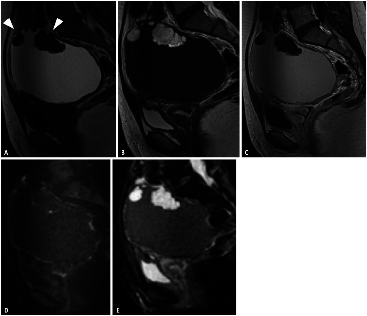
Fig. 5. Polypoid endometriosis in a 24-year-old female with dysmenorrhea and lower abdominal pain.
The patient received cervical cancer screening. An ovarian tumor is found.
A-E. Sagittal T1WI (A); sagittal T2WI (B); sagittal contrast-enhanced T1WI (C); diffusion weighted image (D); apparent diffusion coefficient map (E). Tumor contents showed high signal intensity on T1WI and strong low signal intensity on T2, which indicates a hemorrhagic cyst. The cyst wall is thick. These findings indicate an endometriotic cyst. Multiple mural nodules (arrowheads) show a high signal intensity on T2WI. Each nodule includes many septations on T2WI. Contrast-enhanced T1WI shows no solid lesions within the mural nodules. No area has restricted diffusion. WI = weighted image

