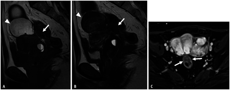
Fig. 6. Rectal endometriosis in a 33-year-old female with dysmenorrhea.
A, B. Sagittal T1WI (A); sagittal T2WI (B). Ovarian endometriotic cyst (arrowheads), which shows high signal intensity on T1WI (A); shading on T2WI (B) is apparent on the uterine body. The findings of an elevated posterior vaginal fornix, tethered appearance of the rectum to the uterus, and a fibrotic plaque covering the serosal surface of the uterus indicates Douglas obliteration. Thickening of the rectal anterior wall of the rectum showing a mushroom cap sign (arrows) is observed at a height of 16 mm. This finding indicates rectal endometriosis. C. On axial contrast-enhanced T1WI, rectal endometriosis is enhanced. It involves more than half of the circumference (arrows). WI = weighted image

