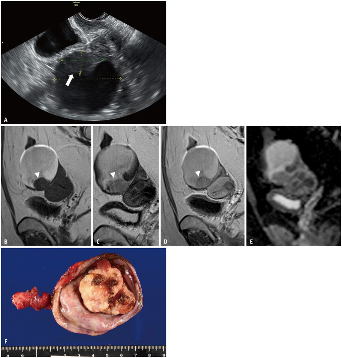
Fig. 7. Four years after Figure 6; the patient had taken progestins to treat her endometriosis.
A. Upon follow-up examination of the ovarian endometriotic cyst (depicted in Fig. 6) by transvaginal ultrasonography, the mural nodule (arrow) has emerged within the cyst. B-E. Sagittal T1WI (B); sagittal T2WI (C); sagittal contrast-enhanced T1WI (D); ADC map (E). The endometriotic cyst size is similar to that shown in Figure 6, even after hormonal therapy. On T2WI (C), the fluid intensity increased, although T2 dark spot signs are still visible. Mural nodules (arrowheads) are observed at the bottom of the cyst. The nodule (arrowhead) is well-enhanced on contrastenhanced T1WI (D) and there is low signal intensity on the ADC map (E), indicating restricted diffusion. F. Surgery has been performed. An irregularly shaped mural nodule is observed within the cyst. The mass has beem pathologically diagnosed as clear cell carcinoma (FIGO stage Ia). ADC = apparent diffusion coefficient, WI = weighted image

