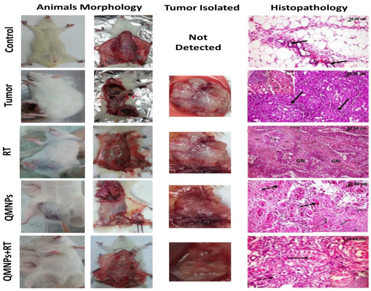
Figure 5.
QMNPs and/or RT effect on morphological and histopathological indices. (Control rat) showing normal mammary acini (arrow) embedded in the surrounding adipose tissue. (Tumor rat) showing many empty spaces (arrow) in the cribriform adenocarcinoma, the top left image is showing adenocarcinoma, notice the multiple spaces within the solid masses of the tumor some are empty and others contained pink proteinaceous secretion (arrow). (RT rat showing large eosinophilic granular areas of cancer cells’ necrosis [GN] among the tumor masses.). (QMNPs rat) showing marked necrosis of the cancer cells with its transformation into hyalinized eosinophilic material (arrow) as well as many pyknotic nuclei. QMNPs + RT rat showing necrosis and beginning of fibrous bands (arrow) among the cancer cell groups. Tumor size was measured using a standard caliber, in length and width, and the tumor was weighed upon excision it of all treated and untreated tumor-bearing rats. Number of animals (5 per group).
QMNP indicates quercetin magnetite nanoparticles; RT, radiotherapy.

