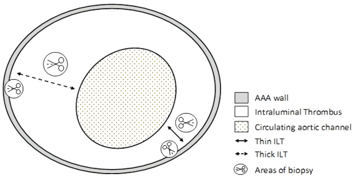
Figure 1.
Transverse view of representative abdominal aortic aneurysm, demonstrating the sites of sampling. Arrows point out areas of biopsy: a thick section with a thickness ≥25 mm; a thin section with a thickness of ≤10 mm plus each of two sections of the wall: one section adjacent to the thick part of the ILT and one section adjacent to the thin part of the ILT.

