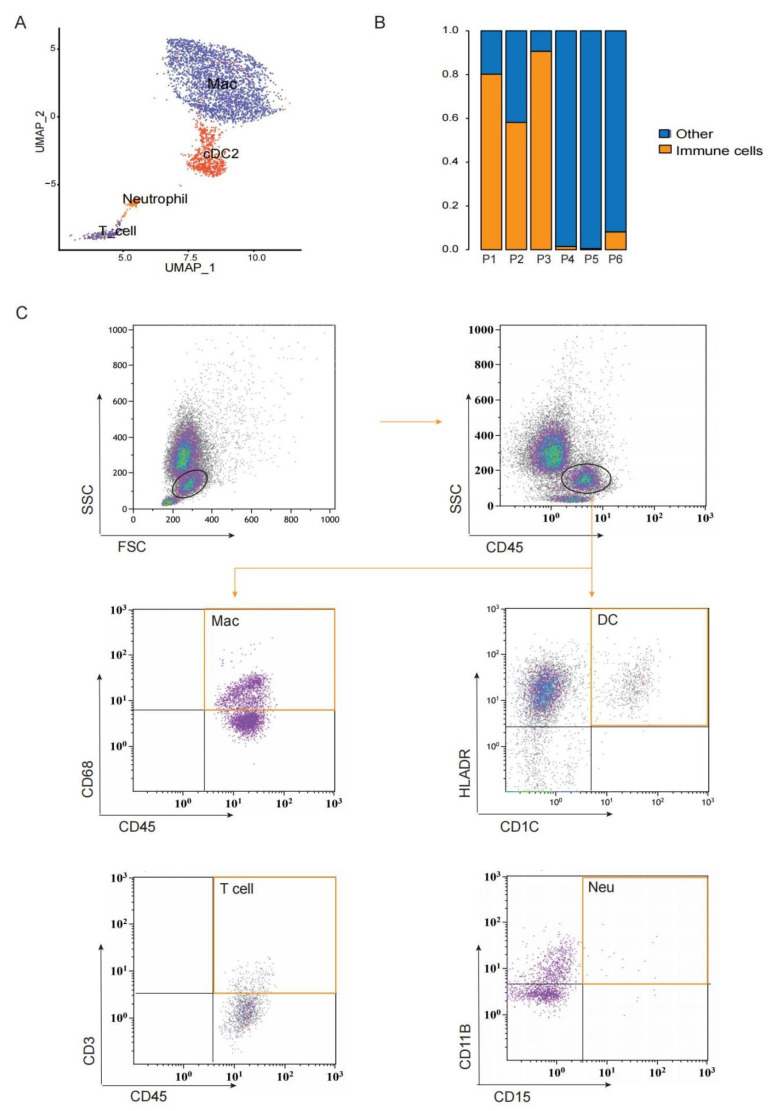
Figure 4.
Immune cells exist in preovulatory follicles. (A) UMAP showing immune cell types in preovulatory follicles. (B) The proportion of immune cells in each preovulatory follicle sample. (C) Flow cytometry analysis identifying CD45+ immune cells, macrophages (sorted with CD45 and CD68), DCs (sorted with CD1C and HLA-DR), T cells (sorted with CD45 and CD3) and neutrophils (sorted with CD11B and CD15) in a single preovulatory follicle.

