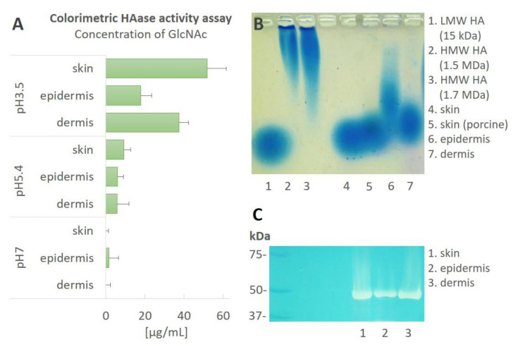
Figure 4.
Hyaluronidase activity in the skin. Hyaluronidase activity was analyzed in the homogenates (10 mg proteins/mL) from the human skin, epidermis and dermis. (A) Colorimetric hyaluronidase activity assays of the samples incubated at differing pH values (pH 3.5, pH 5.4 and pH 7). The values represent the means of three independent biological samples ± the SEM of the GlcNAc concentrations. (B) HA size analysis via agarose gel electrophoresis at pH 3.5. The samples were incubated with exogenous HMW HA (1.5 MDa) at 37 °C for 24 h. The samples were then analyzed via agarose gel electrophoresis and HA staining (blue). The LMW HA and both HMW HA served as the size references. (C) A representative zymography using HMW HA as a substrate. The left-most lane shows a protein ladder. Abbreviations: low molecular hyaluronan (LMW HA), high molecular hyaluronan (HMW HA), N-acetylglucosamine (GlcNAc).

