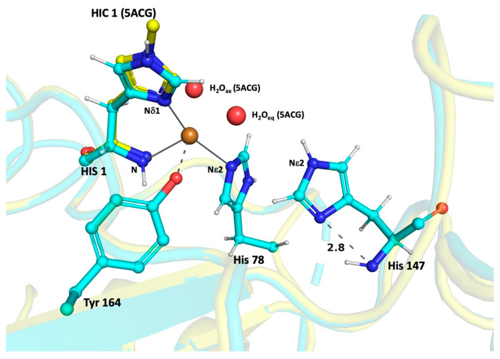
Figure 4.
Results from His-only unrestrained refinement in LsAA9AEc_2 structure (cyan colored). His147 shows single protonation Nε2 atom. The Nis-brace residues (internal control) show their single protonation state. The H-bond distance between the Nδ1 and backbone N atoms of His147 is shown in Å. The structure of LsAA9A_Ao (PDB id: 5ACG, yellow colored) is shown for comparison with 1st His methylated (HIC) and the two water molecules bound to the Cu(II). The oxygen species binds at the position of H2Oeq. The figure was prepared with Pymol.

