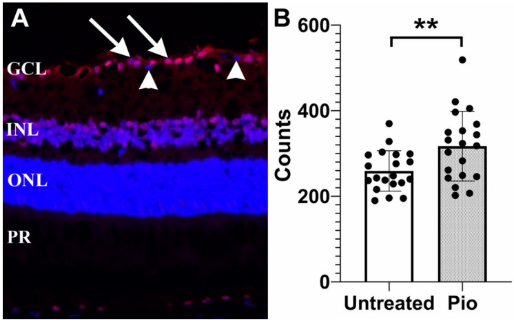
Figure 1.
Protective effect of pioglitazone in DBA/2J mice. (A) Representative image showing γ-synuclein labeling of RGCs (arrows). Displaced amacrine cells are not bound by this antibody and are not counted (arrowheads) (B) RGC density in pioglitazone treated mice (Pio) is significantly higher than in untreated controls (p = 0.009). Each dot represents one eye. GCL: Ganglion cell layer; INL: Inner nuclear layer, ONL: Outer nuclear layer, PR: Photoreceptor inner segments. **: p < 0.01.

