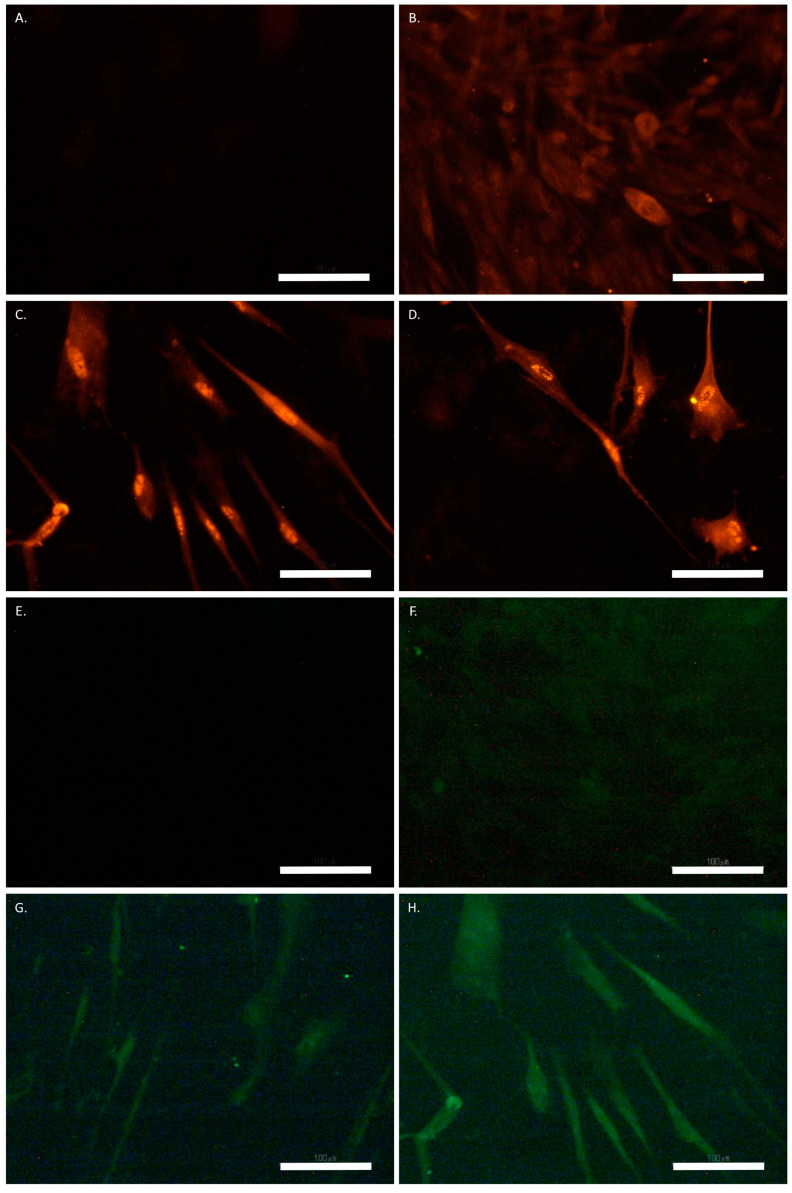
Figure 7.
SCAP treated with UDP-4 and stained for GFAP (red) and NeuN (green). (A) Untreated cells stained with the secondary antibody for GFAP-isotype control. (B) Untreated cells stained for GFAP, showing basal expression. (C) UDP-4-treated cells after 3 weeks, showing medium expression of GFAP. (D) UDP-4-treated cells after 4 weeks, showing strong expression of GFAP. (E) Untreated cells stained with the secondary antibody for NeuN-isotype control. (F) Untreated cells stained for NeuN, showing no expression. (G) UDP-4-treated cells after 2 weeks, showing basal expression of NeuN. (H) UDP-4-treated cells after 3 weeks, showing medium NeuN expression. Experiments were conducted in duplicates. Scale bars correspond to 100 µm.

