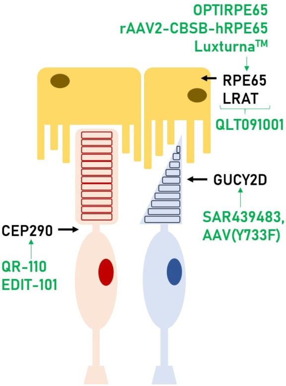
Figure 1.

Photoreceptor (rod in red, cone in blue) and RPE cells. In this scheme, we can see where each gene discussed in this review has its function within the retina. These genes are present in both rods and cones, however, we depicted it showing the cell type where relative expression is highest. Also, in green, the therapies that are currently available or under investigation for those genes. RPE, retinal pigment epithelium.
