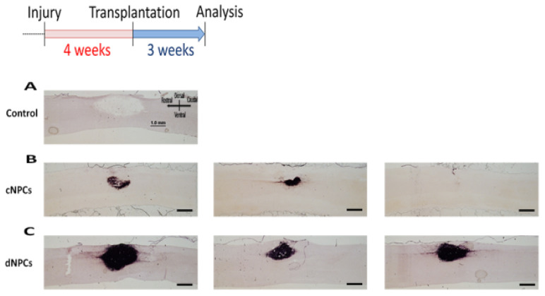
Figure 2.
NPC transplants survival. Two types of cells were injected into the lesion site 4 weeks after injury and survived for 3 weeks after transplantation. AP histochemical staining demonstrated that there was no AP+ staining in the control group (A). In cNPCs group, transplanted cells did not show good survival with some AP+ cells inside the lesion but not filling the lesion area and some animals with no AP+ cells inside the lesion/transplant area (B). In dNPCs group, transplanted cells survived in all rats and filled the lesion/transplant area (C). Scale bar = 1.0 mm.

