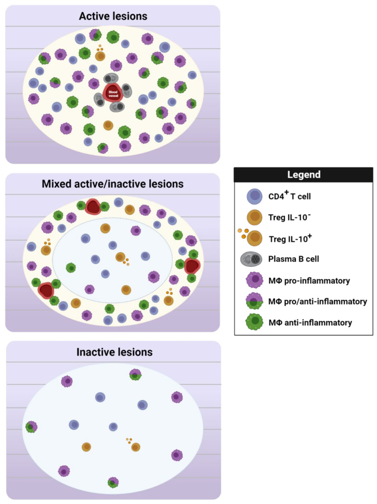
Figure 2.
Scheme of the regulatory cells present in MS lesions. The IL-10+ Treg/Treg ratio increases in the plaque of active and inactive lesions and at the rim of mixed active/inactive areas (Zandee et al., 2017). Perivascular CD138+CD38+ plasma cells act as a source of the anti-inflammatory cytokine IL-10, and they are mainly observed in active lesions (Machado-Santos et al., 2018). In terms of myeloid cells, the distribution of typical markers used to identify pro- or anti-inflammatory cells in demyelinating lesions reveals the presence of myeloid cells with an intermediate activation phenotype (CD40+/CD206+ cells) in active lesions (Vogel et al., 2013) and the coexistence of pro-inflammatory and regulatory myeloid cells at the rim of both mixed active/inactive and inactive lesions (Vogel et al., 2013; Jackle et al., 2020). However, anti-inflammatory myeloid cells are also observed in active lesions and at the rim of mixed active/inactive lesions (Miron et al., 2013). Legend: CD4+ T cell = CD4+ FoxP3−; Treg IL-10− = CD4+ FoxP3+IL-10−; Treg IL-10+ = CD4+ FoxP3+ IL-10+; plasma B cell = CD138+CD38+; Mϕ pro-inflammatory = iNOS+ or CD40+ cells; Mϕ pro-/anti-inflammatory = CD40+CD206+ cells; Mϕ anti-inflammatory = CD206+ or CD163+ cells. Figure created with Biorender.com (accessed on 12 January 2022).

