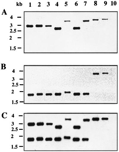
FIG. 5.
Autoradiogram after Southern hybridizations of C. meningosepticum genomic DNAs. The DNAs were restricted with XmnI, and the probe consisted of the 372-bp DraI-XmnI fragment (S1) of pBS1 (A), the 306-bp XmnI-DraI fragment (S2) of pBS1 (B), or the 667-bp DraI fragment (S3) of pBS1 (C) (see Fig. 1 for the locations of S1, S2, and S3). Lanes: 1, C. meningosepticum PINT; 2, C. meningosepticum CIP 6058; 3, C. meningosepticum AMA; 4, C. meningosepticum GEO; 5, C. meningosepticum CIP 7830; 6, C. meningosepticum CIP 6059; 7, C. meningosepticum CIP 7905; 8, C. meningosepticum AB 1572; 9, C. meningosepticum H01J100; 10, E. coli DH10B (negative control). The sizes of the DNA fragments may be deduced from the scale (1.5 to 4 kb) shown on the left sides of the gels.

