Fig. 3.
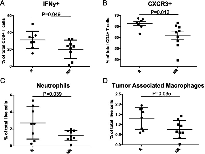
Responder colonized mice show anti-tumor immune phenotype following treatment with immunotherapy. Half of the resected subcutaneous allograft tumors from human microbiota colonized responder and non-responder mice (n = 9/group) were subjected to single-cell dissociation and flow cytometric analysis for A tumoral CD8+ IFNγ+ T cells as represented by percent of total intra-tumoral CD8+ T cells in responder (n = 8) and non-responder (n = 9) tumors, Mann-Whitney P = 0.049; B tumoral CD4+ CXCR3+ T cells as represented by percent of total CD4+ T cells in responder (n = 7) and non-responder (n = 9) tumors, Mann-Whitney P = 0.012; C tumoral neutrophils (Gr1+ CD11c+ CD11b+ cells) as represented by percent of total live cells in responder (n = 8) and non-responder (n = 9) tumors, Mann-Whitney P = 0.039; and D tumoral macrophages (Gr1− CD11c− CD11b+ cells) as represented by percent of total live cells in responder (n = 8) and non-responder (n = 9) tumors, Mann-Whitney P = 0.035

