Fig. 4.
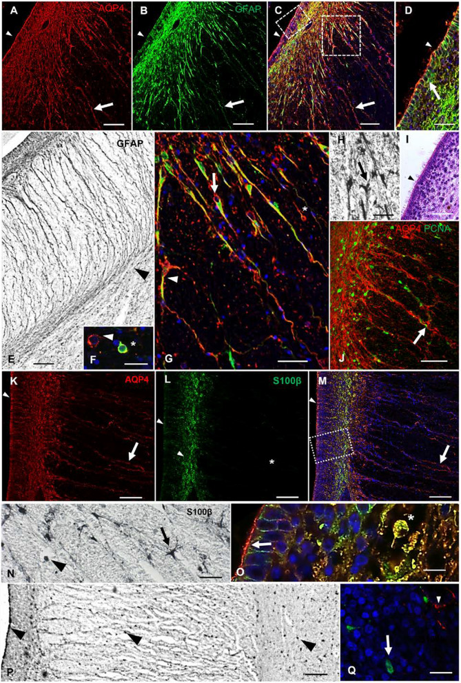
AQP4 and astrocytic markers in a control sample of 21 PCW. a AQP4 immunofluorescence. The arrow indicates immunodetection of AQP4 in cellular processes, while the arrowhead indicates immunodetection of AQP4 at the apical poles of ependymal cells. b Anti-GFAP immunofluorescence. Arrow indicates a positive GFAP radial process; arrowhead indicates the absence of GFAP at the apical pole of the ependyma. c Merge between a and b. Arrow indicates colocalization in cellular processes. Arrowhead indicates immunodetection of AQP4 at the apical pole of the ependyma. d Magnification of square in c showing VZ. Arrow points to positive GFAP immunostaining on basolateral walls, while AQP4 mostly labels the apical pole of the ependyma (arrowhead). e GFAP immunohistochemistry of the fornix region, showing GFAP staining in basal processes that curve to follow the fiber tracts of the fornix (arrowhead). f Detail of cells with intermediate progenitor morphology. Arrowhead points to AQP4 expression. Asterisks marks colocalization of AQP4 and GFAP. g Magnification of the subventricular zone in C. Arrow points to basal radial glial cells with AQP4 staining only in the cell body, but colocalized AQP4 and GFAP staining in the cell process. Arrowhead point to an AQP4-positive cell that is differentiating into an astrocyte. Asterisk marks a cell with basal radial glia morphology emitting a cellular process towards the apical zone. h Detail of the subventricular area near the fornix immunostained with anti-GFAP. Arrow points to an astrocyte. i Hematoxylin–eosin staining of a section stained in parallel with d Arrowhead points to characteristic multicilia on ependymal cells. j Immunolabeling against AQP4 and PCNA (mitosis marker) in a section stained in parallel with g. AQP4-positive cells are restricted to the white matter tracts and serve as a scaffold for proliferative cells. Arrow indicates a proliferative cell migrating through a radial process. k Anti-AQP4 immunofluorescence in a slide stained in parallel with a. Arrow indicates immunodetection of AQP4 in cellular processes. Arrowhead points to immunodetection of AQP4 at the apical pole of the neuroepithelium. l Anti-S100β immunofluorescence. Arrowheads point to the expression of S100β in the ependymal and subventricular cells. The asterisk indicates lack of S100β expression in the radial processes. m Merge between k and l. Arrow points to AQP4 staining in cellular processes. Arrowhead points to immunodetection of AQP4 at the apical pole of the ependyma. n Detail of S100ß expression in the subventricular zone. Arrow points to a differentiated astrocyte and arrowhead indicates an undifferentiated cell. o Magnification of the VZ in M. Arrow points to positive cytoplasmic immunostaining for S100β. Asterisk marks colocalization in the VZ. p Panoramic view of the medial parieto-occipital cortex labeled with anti-S100β, where the absence of radial processes is observed. Arrowheads indicate expression in cellular bodies of the ependymal cells and throughout the parenchyma. q Slide stained in parallel with P and double labeled with AQP4 (red) and S100β (green) at the cortical plate area, showing no colocalization between AQP4 (arrowhead) and S100ß (arrow). PCW, post-conceptional weeks; VZ, ventricular zone. Scale bar: a, b, c, i 200 µm; d, g, h, n, q, 40 µm; e, p 100 µm; f, o 20 µm; j 50 µm; k–m 80 µm

