Fig. 5.
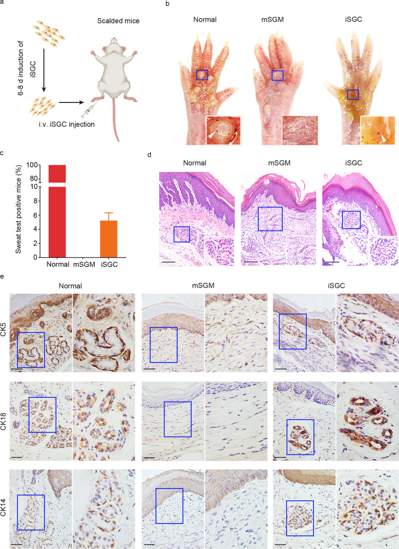
Engraftment of iSGC functionally restored SG. a Schematic diagram representing the experimental procedure. b Starch-iodine sweat tests on paw skin of thermal-injured mice showed that paw pads of mSGM-treated mice (n = 60), iSGC-treated mice (n = 60) and normal mice (n = 60) responded by displaying indigo-black dots on day 21 after transplantation. c The positive rate of sweat test of the iSGC-treated group, (5.2 ± 1.1)% of the recipient mice (n = 60). d H&E staining was conducted to visualize normal group and mSGM- and iSGC-treated wounds on day 21 post-injury. Emerging glandular structures were seen in the dermis of iSGC-treated mice and normal mice. Scale bar = 50 μm. Illustrations, higher magnification of the boxed areas. e The SG markers CK5, CK18, and CK14 were assessed by immunohistochemical analysis to examine the SG formation. The results showed that, like natural paw shin, the iSGC-treated group could form SG-like structures with positive staining for the SG markers, while no SG regeneration was observed in mSGM-treated group. Scale bars = 50 μm. Illustrations, higher magnification of the boxed areas. iSGC induced sweat gland-like cell, SG sweat gland, mSGM modified sweat gland culture medium, CK5 cytokeratin 5, CK18 cytokeratin 18, CK14 cytokeratin 14

