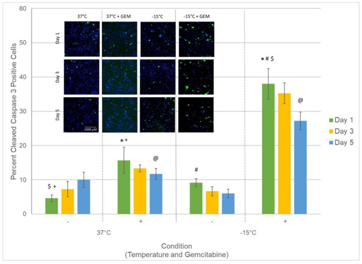
Figure 6.
Analysis of percent of PaCa cells with active caspase-3 following treatment. Samples were treated with freezing to −15 °C, 50 nM gemcitabine, or the combination. The percent of cells with cleaved caspase-3 (active caspase-3) present within the nucleus was assessed at 1, 3, and 5 days post-treatment to determine the level of involvement and timing of apoptotic cell death associated with each treatment. Data reveal a significant increase in the number of cells with cleaved caspase-3 in gemcitabine/−15 °C combination samples compared to either treatment alone. The increased and prolonged nuclear presence of active caspase-3 indicates increased apoptotic activity in combination samples, which is believed to be responsible for the observed increased cell death. Insert: Representative mosaic fluorescent micrographs, acquired using the CX5 high throughput image analysis system, of the remaining cells (blue) and subpopulation of cells with active caspase-3 within the nucleus (green) (Scale Bar = 2000 µM). Data in bar graph represent the average (±SD) of the analysis of the mosaic images acquired from 9 replicate samples. (@, #, $, *, + = p < 0.01).

