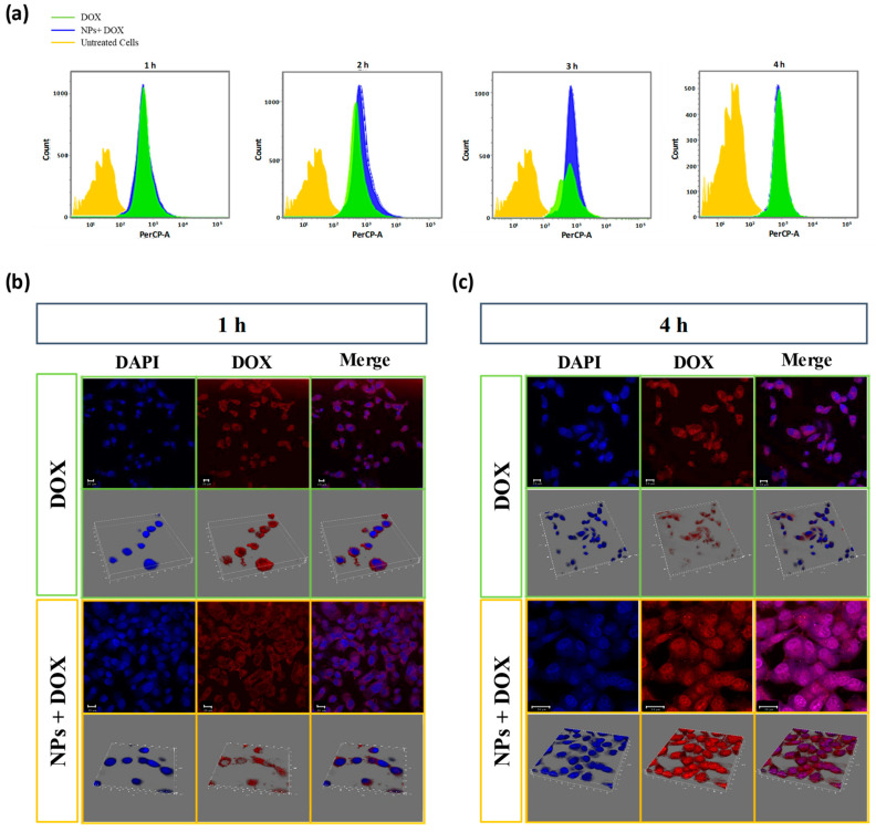
Figure 5.
(a) Flow cytometry analysis of cellular uptake of α1-acid glycoprotein-conjugated hyaluronic acid nanoparticles (AGP-HA NPs) + DOX and free DOX in MDA-MB-231 cells incubated for 1, 2, 3, and 4 h. (b,c) CLSM images of α1-acid glycoprotein-conjugated hyaluronic acid nanoparticles (AGP-HA NPs) + DOX and free DOX in MDA-MB-231 cells incubated for 1 h (b) and 4 h (c). Nuclei were stained in blue with DAPI dyes, and DOX fluorescence in cells is red.

