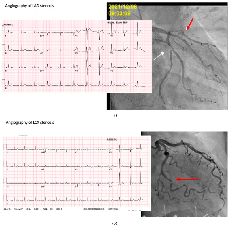
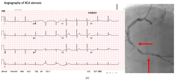
Figure 4.
ECG examples of the AI model predicting the coronary lesions, arrows indicate stenosis by coronary angiography result. (a) ECG of a patient with >70% stenosis in LAD and about 30% stenosis in LCX in angiography. (b) ECG of a patient with >70% stenosis in LCX in angiography. (c) ECG of a patient with >70% stenosis in RCA in angiography.


