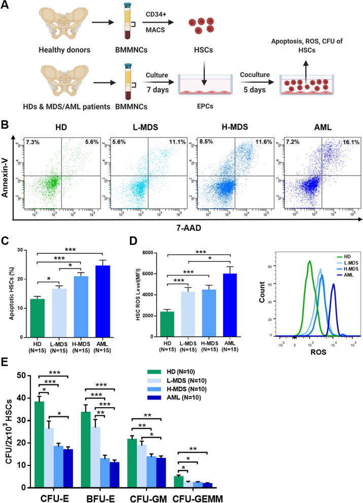
Fig. 3.
Supporting abilities of BM EPCs from MDS patients to HSCs. A Schematic diagram of the study design for the BM EPC coculture process with HSCs. After 5 days of coculture, the apoptosis ratio and the quantification of the level of intracellular ROS and CFU efficiencies of HSCs were detected. Representative images (B) and quantification (C) of the apoptosis ratio of HSCs after coculture with BM EPCs are shown. Quantification (D, left) and representative images (D, right) of the ROS levels (MFI) of HSCs after coculture are shown. E The CFU plating efficiencies of HSCs, including CFU-E, BFU-E, CFU-GM and CFU-GEMM, after coculture with BM EPCs from HDs, L-MDS, H-MDS and AML patients. Statistical analyses were performed using the Mann–Whitney U test. Data are presented as the means ± SEM (*P ≤ 0.05, ** P ≤ 0.005, *** P ≤ 0.001). AML Acute myeloid leukaemia, BFU-E Burst-forming unit erythroid, BM Bone marrow, BMMNCs Bone marrow mononuclear cells, CFU Colony-forming unit, CFU-E Colony-forming unit erythroid, CFU-GM Colony-forming unit-granulocyte/macrophages, CFU-GEMM Colony-forming unit-granulocyte, erythroid, macrophage and megakaryocyte, EPCs Endothelial progenitor cells, HD Healthy donor, HSC haematopoietic stem cells, H-MDS Higher-risk myelodysplastic syndromes, L-MDS Lower-risk myelodysplastic syndromes

