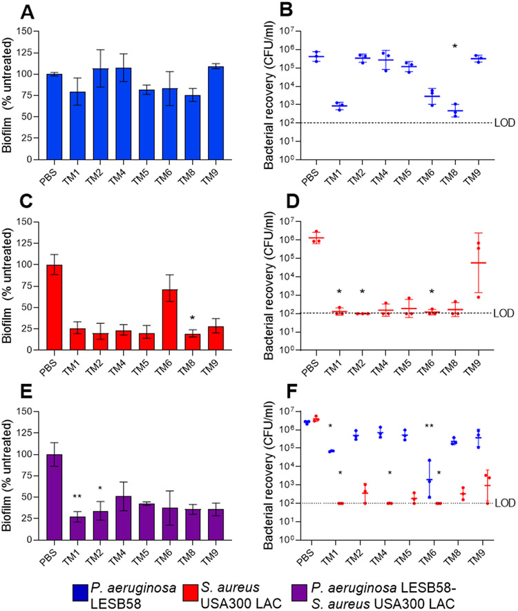
Figure 6.
Effect of peptoids on mono- and polymicrobial biofilms. Peptoids (31.25 μg/ml) were used to treat biofilms comprising (A,B) P. aeruginosa LESB58 (5 × 10⁵ CFU/ml), (C,D) S. aureus USA300 LAC (2.5 × 10⁷ CFU/ml) or (E,F) both species. Biofilms were grown for 20-24 h in DMEM-FBS-G prior to treatment and re-incubated for another 20-24 h. (A,C,E) Biofilm was quantified by CV staining (%) and (B,D,F) and bacterial recovery from biofilms (CFU/ml) determined on selective agar plates. The dotted line indicates the limit of detection (LOD) at 102 CFU. Data from three independent experiments each (n = 3) are shown as (A,C) mean ± SEM or geometric mean ± geometric SD. * P < 0.05, ** P < 0.01 according to Kruskal-Wallis test with Dunn’s correction.

