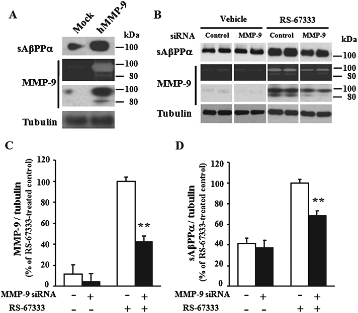
Fig. 3.

sAβPPα production by MMP-9 induction. A) H4/AβPP cells transfected with MMP-9 or mock plasmid were maintained for 48 h in a serum-free medium. The concentrated medium was analyzed by gelatin zymography and immunoblotting using anti-MMP-9 or sAβPPα antibodies. B) H4/AβPP/5-HT4 cells transfected with MMP-9 or control siRNA were cultured for 2 days and then treated with serum-free medium or RS-67333 (3 μM) for 24 h. The concentrated and immunoprecipitated medium were analyzed by the gelatin zymography and immunoblotting using anti-MMP-9 or sAβPPα antibody, respectively. The tubulin band from the cell lysate was used as an internal control. Band intensities of immunoreactive MMP-9 (C) and sAβPPα (D) were quantified, and the percent intensity normalized with tubulin was indicated. Error bars show S.E.M. (n = 4). **p < 0.01.
