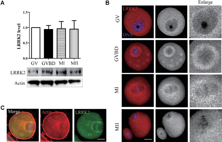
Figure 1.
Expression and localization of LRRK2 in mouse oocyte meiosis. (A) The expression level of LRRK2 at different stages (GV, GVBD, MI, and MII) was examined by western blotting in mouse oocytes. (B) The localization of LRRK2 was certificated by LRRK2 antibody in mouse oocytes. LRRK2 dispersed in the cytoplasm at the GV stage and was mainly distributed around the spindle after GVBD. Red, LRRK2; blue, DNA; scale bar, 20 μm. (C) LRRK2 was colocalized with actin around the spindle in the MI-stage oocytes. Red, actin; green, LRRK2; blue, DNA; scale bar, 20 μm.

