Figure 5.
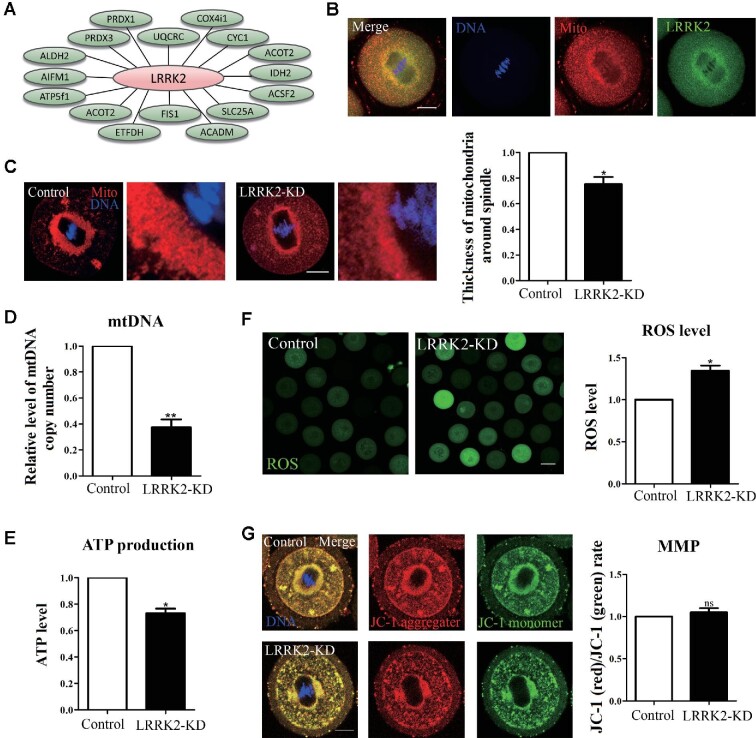
LRRK2 depletion affects the distribution and function of mitochondria in mouse oocytes. (A) Screening mitochondria-related proteins associated with LRRK2 by MS analysis. (B) LRRK2 showed a similar localization pattern to mitochondria in mouse oocytes. Red, mitochondria; green, LRRK2; blue, DNA; scale bar, 20 μm. (C) The accumulation of mitochondria around the spindle in the LRRK2 siRNA-injected oocytes was decreased compared with that in the control oocytes. Red, mitochondria; blue, DNA; scale bar, 20 μm. (D) The relative content of mtDNA copy number in the LRRK2 siRNA-injected oocytes was significantly decreased compared with that in the control oocytes. (E) The relative content of ATP in the LRRK2 siRNA-injected oocytes was significantly decreased compared with that in the control oocytes. (F) The relative fluorescence intensity of ROS in the LRRK2 siRNA-injected oocytes was significantly increased compared with that in the control oocytes. Scale bar, 50 μm. (G) JC-1 staining showed no significant difference in MMP between the LRRK2 siRNA-injected and control oocytes. Red, JC-1 aggregates; green, JC-1 monomer; blue, DNA; scale bar, 20 μm. *P < 0.05, **P < 0.01, and ns, no significant difference.

