Figure 3.
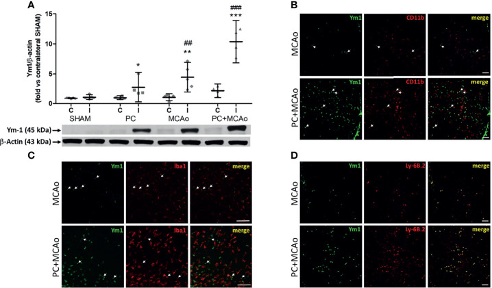
Ischemic PC potentiates the elevation of the M2/N2 marker Ym1 caused by 1h MCAo in myeloid cells of the ischemic cortex of mice. (A) Western blotting analysis of Ym1 expression in tissue homogenates from the contralateral (C) and ipsilateral (ischemic, I) cortex of mice subjected to SHAM surgery, ischemic PC (15 min MCAo followed by 72h of reperfusion), 1h MCAo followed by 24h of reperfusion (MCAo) or PC+MCAo. *P = 0.0479, **P = 0.0079 and ***P = 0.0004 vs corresponding contralateral; ##P = 0.0059 and ###P = 0.0003 vs ipsilateral SHAM (two-way ANOVA followed by Bonferroni post-test, n=4-5 mice per experimental group). Representative immunofluoresce images showing Ym1 expression (green fluorescence) in CD11b immunopositive myeloid cells [red fluorescence in (B)], Iba1 immunopositive microglia/macrophages [red fluorescence in (C)] or Ly-6B.2 immunopositive myeloid cells [granulocytes and monocytes/macrophages, red fluorescence in (D)] in the cortex of mice undergone 1h MCAo preceded (72h before) by sham surgery (MCAo) or by ischemic PC (PC+MCAo). Arrows indicate co-localization of Ym1 with CD11b or Iba1 in amoeboid-shaped cells. Scale bars = 100 μm.

