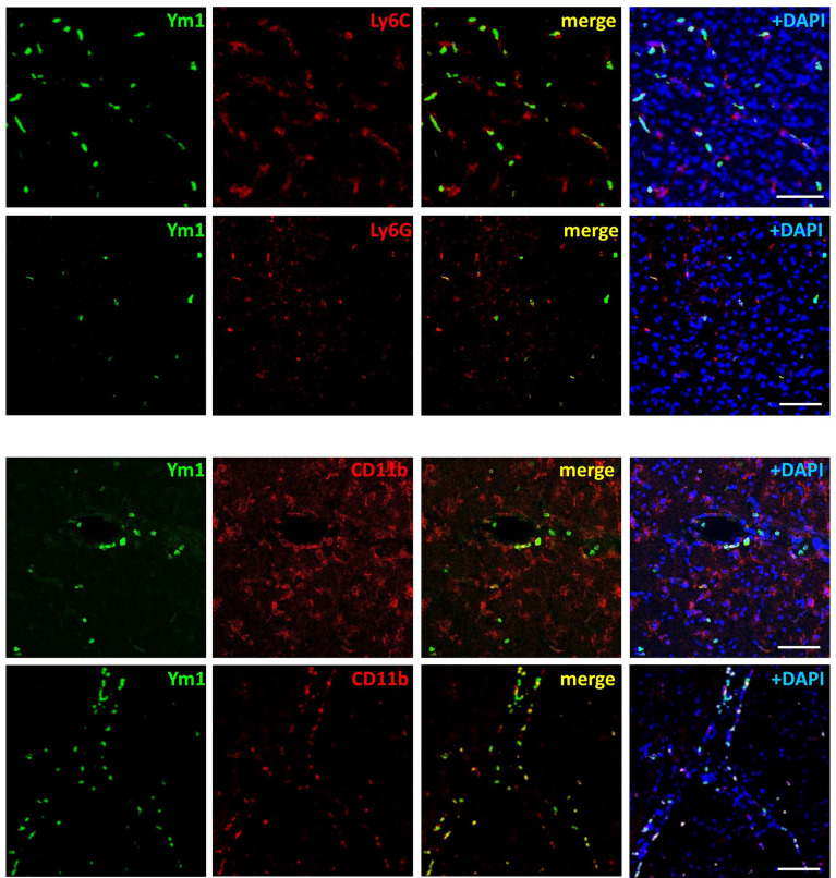
Figure 4.
Ym1 is expressed in myeloid cells infiltrating from the periphery, resembling monocytes/macrophages and neutrophils. Representative immunofluorescence images showing Ym1 expression (green fluorescence) in CD11b immunopositive myeloid cells (lining the blood vessels and populating the perivascular space), in Ly-6C immunopositive monocytes/macrophages and in Ly-6G immunopositive neutrophils. Nuclei are counterstained with DAPI (blue signal). Scale bars = 50 μm.

