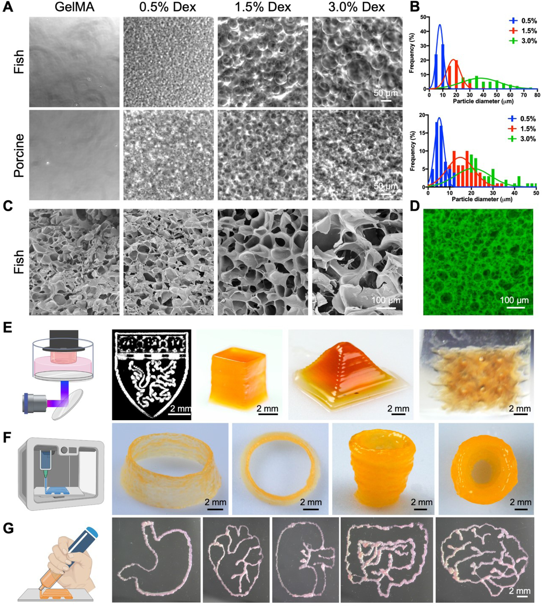
Figure 2.

Characterizations and bioprinting of the PGelDex bioinks. (A) Optical micrographs showing the fish- and porcine-derived PGelDex bioinks containing different dextran concentrations (0.5, 1.5, and 3.0 wt.%). (B) Quantification data showing the size distributions of the dextran emulsion droplets of the PGelDex bioinks at different dextran concentrations (0.5, 1.5, and 3.0 wt.%). (C) SEM images showing the interconnected porous structures at dextran concentrations of 0.5, 1.5, and 3.0 wt.%. (D) Confocal fluorescence micrograph showing the interconnected porous structures in the construct with 3.0 wt.% of dextran. (E-G) 2D and 3D constructs fabricated with (E) DLP bioprinting, (F) extrusion bioprinting, and (G) handheld bioprinting using the PGelDex bioinks. Part of the figure drawn with BioRender.
