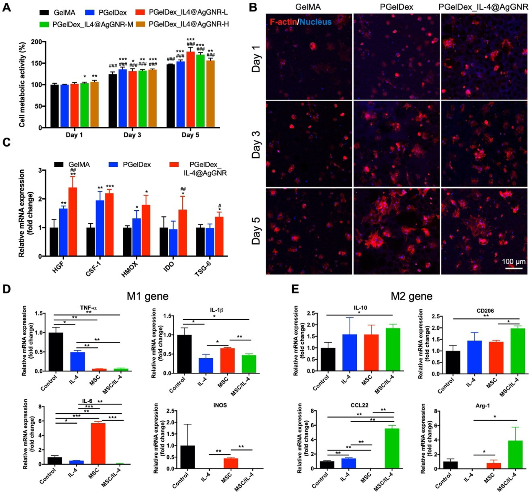
Figure 5.

MSC- and IL-4@AgGNR-incorporated PGelDex micropore-forming bioink polarized macrophages into an anti-inflammatory phenotype. (A) Quantified cytocompatibility evaluations of MSCs cultured in the constructs bioprinted with GelMA, PGelDex, and PGelDex with different IL-4@AgGNR concentrations. (B) Fluorescence micrographs showing bioprinted MSCs in GelMA, PGelDex, and IL-4@AgGNR-incorporated PGelDex samples on days 1, 3, and 5 of culture. (C) Gene expressions of MSCs encapsulated in GelMA, PGelDex, and IL-4@AgGNR-incorporated PGelDex constructs normalized to reference gene RPL13A in GelMA. (D, E) Representative macrophage phenotype markers of THP-1 cells cultured in PGelDex, IL-4@AgGNR-incorporated PGelDex, MSC-encapsulated PGelDex, and MSC-IL-4@AgGNR-incorporated PGelDex constructs through a transwell assay. Relative mRNA expressions were normalized to reference gene GAPDH in GelMA. *P < 0.05, **P < 0.01, ***P < 0.001; one-way ANOVA (A and C, compared with the GelMA control group; D and E, compared with the PGelDex control group); ###P < 0.001; one-way ANOVA (A, compared with the corresponding groups on day 1; C, compared with the PGelDex group); n = 3.
