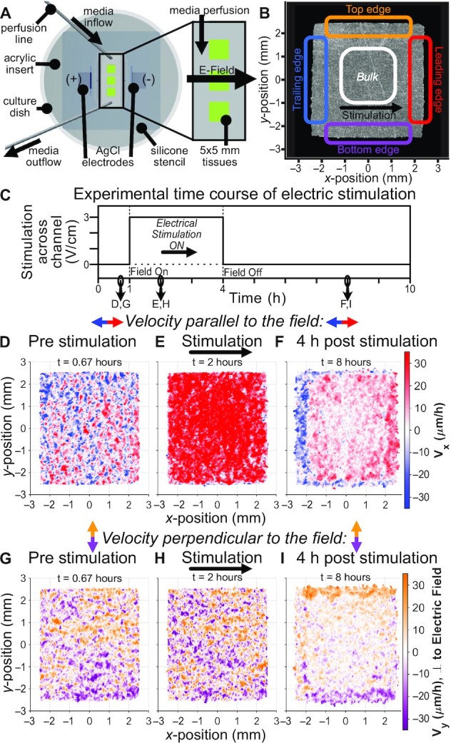
Fig. 1.

Collective electrotaxis step response induces a spatiotemporal effect in epithelial tissues persisting long after stimulation. (A) Diagram of electro-bioreactor providing electrical stimulation in the x-axis and perfusion of fresh media in the y-axis through the chamber containing three 5 × 5 mm tissues. (B) Phase contrast image of a single MDCK epithelial tissue; annotations denote specific regions analyzed throughout the paper. (C) Trace of electrical stimulation step response stimulus: 1 h unstimulated, 3 h ‘rightward’ stimulation, and 6 h poststimulation. (D)–(F) Single tissue heatmaps of velocity in the x-direction, aligned with the direction of electric field (red). Note that (E) indicates large increase in the rightward collective motion during stimulation, while (F) indicates a new relaxation behavior hours after stimulation. See Figure S1 (Supplementary Material). for corresponding control tissue heatmaps. (G)–(I) Analogous heatmaps of velocity in the y-axis, perpendicular to the direction of electric field. Note again the new pattern long after stimulation ended in (I), despite minimal apparent changes during stimulation in (H).
