. 2022 Jan 11;303(1):215–225. doi: 10.1148/radiol.211028

Figure 3:

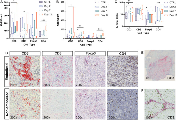
Transarterial embolization induces increased lymphocyte infiltration in embolized tumors. Box and whisker plots show (A) the average number of CD3+, CD8+, FOXP3+, and CD4+ cells (intrastromal and intratumoral cells) at 2 days (n = 7), 7 days (n = 12), and 12 days (n = 17) after embolization relative to control (CTRL) tumors that were not embolized (n = 12); (B) the average number of cells within the intratumoral compartment of embolized tumors at 2, 7, and 12 days after embolization relative to control tumors that were not embolized; and (C) the average percentage of immune cells found within the intratumoral compartment expressed as a percentage of total number of cells (intrastromal plus intratumoral cells) at 2, 7, and 12 days after embolization relative to control tumors that were not embolized. Whiskers indicate maximum and minimum values. Boxes extend from the 25th to 75th percentiles. The line in the middle of the box is plotted at the median. * = P < .05, ** = P < .01, and *** = P < .001 according to the generalized estimating equation model. Significant P values (left to right): in A, .02, .03, .01, <.001; in B, .02, .02, .001, <.001; and in C, .02, .007, .01, .002. (D) Representative images of histologic staining of CD3, CD8, FOXP3, and CD4 cell markers in embolized and control tumors. (E) Representative low-magnification image shows CD3+ cell infiltration into a hepatocellular carcinoma (HCC) tumor 12 days after embolization. (F) Representative image demonstrates intrastromal CD3+ cells (pink markers) and intratumoral CD3+ cells (green markers) within HCC specimens.

Transarterial embolization induces increased lymphocyte infiltration in embolized tumors. Box and whisker plots show (A) the average number of CD3+, CD8+, FOXP3+, and CD4+ cells (intrastromal and intratumoral cells) at 2 days (n = 7), 7 days (n = 12), and 12 days (n = 17) after embolization relative to control (CTRL) tumors that were not embolized (n = 12); (B) the average number of cells within the intratumoral compartment of embolized tumors at 2, 7, and 12 days after embolization relative to control tumors that were not embolized; and (C) the average percentage of immune cells found within the intratumoral compartment expressed as a percentage of total number of cells (intrastromal plus intratumoral cells) at 2, 7, and 12 days after embolization relative to control tumors that were not embolized. Whiskers indicate maximum and minimum values. Boxes extend from the 25th to 75th percentiles. The line in the middle of the box is plotted at the median. * = P < .05, ** = P < .01, and *** = P < .001 according to the generalized estimating equation model. Significant P values (left to right): in A, .02, .03, .01, <.001; in B, .02, .02, .001, <.001; and in C, .02, .007, .01, .002. (D) Representative images of histologic staining of CD3, CD8, FOXP3, and CD4 cell markers in embolized and control tumors. (E) Representative low-magnification image shows CD3+ cell infiltration into a hepatocellular carcinoma (HCC) tumor 12 days after embolization. (F) Representative image demonstrates intrastromal CD3+ cells (pink markers) and intratumoral CD3+ cells (green markers) within HCC specimens.