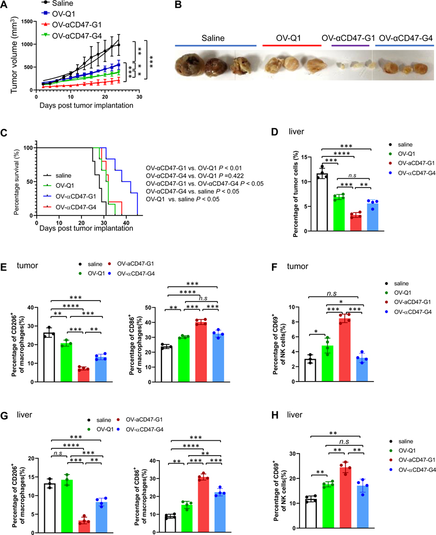
Figure 4. OV-αCD47-G1 improves the therapeutic efficacy against ovarian tumor.
An orthotopic model of human ovarian tumor was established by s.c. injection of 5 × 10⁶ A2780 cells. Two days later, mice were intratumorally injected with a vehicle control or 1 × 10⁵ PFU of OV-Q1, OV-αCD47-G1, or OV-αCD47-G4. (A) Tumor volume of ovarian tumor growth in mice with indicated treatments. (B) The representative tumor images from the four treatment groups. An immunocompetent ovarian cancer metastasis mouse model was established by i.p. injection of 2 × 10⁶ ID8-hCD47 cells expressing a firefly luciferase gene and a GFP gene, following by injected with a vehicle control or 1 × 10⁶ PFU of OV-Q1, OV-αCD47-G1, or OV-αCD47-G4. (C) Survival curve of immunocompetent ovarian cancer mice with indicated treatments (n = 6 each group). Survival was estimated by the Kaplan–Meier method and compared by log-rank test (n=6 mice each group). (D) Percentages of tumor cells among total live cells in the liver. (E) M1 and M2 types of macrophages in tumors from the abdomen were measured by flow cytometry. (F) Percentages of CD69+ NK cells in tumors in the abdomen were measured by flow cytometry. (G-H) Percentages of M1 and M2 types of macrophages (G) and CD69+ NK cells among lymphocyte cells (H) in the liver were measured by flow cytometry. For D to H, Statistical analyses were performed by one-way ANOVA with P values corrected for multiple comparisons by Bonferroni method (n = 3 or 4 mice per group). *P ≤ 0.05; **P ≤ 0.01; ***P ≤0.001; ****P ≤ 0.0001; n.s. non-significant. Data were presented as mean values +/− SD.

